Stack Contrast Adjustment Plugin
Authors:
Jan Michalek (michalek at biomed dot cas dot cz)
Martin Capek (capek
at biomed dot cas dot cz)
Jiri Janacek (janacek at
biomed dot cas dot cz)
Source:
Download Stack_Contrast_Adjustment.jar,
change its name to StackContrastAdjustment.zip and uncompress to retrieve the
source code.
Installation:
Drag and drop Stack_Contrast_Adjustment.jar to the
"ImageJ" window, when "Save
Plugin..." dialog appears, we recommend to put the file in the plugins>Stack
folder. Or download the .jar file to the ImageJ>plugins>Stack folder directly and use Help>Refresh
Menus command. The plugin will then be in the Plugins>Stacks
menu item.
Description:
Fluorescent images captured by a confocal laser
scanning microscope (CLSM) from deep layers of a specimen are often darker than
images from the top layers due to absorption and scattering of both excitation
and fluorescent light. These effects cause problems in subsequent analysis of
biological objects. The plugin implements an algorithm for brightness matching
of CLSM image stacks, based on aligning distribution functions of image pairs.
Prior processing the user should choose and
make active by the slider one slice of the stack - an image with high contrast - as a reference image. Contrast and brightness of images in the stack is
adjusted according to this reference image. The reference image stays intact.
This plugin is designed to perform processing
on 8-bit grayscale and RGB images.
Dialog box parameter:
Unmark Is
background black? checkbox if images in the stack are inverted in
intensities, i.e. their background is white.
The dialog shows the number of the active slice
that is used as the reference image.
A PDF document
and sample confocal stacks (human placenta and
rat muscles) are available.
References:
See also: Our plugin PlaneBrightnessAdjustment.
Example:
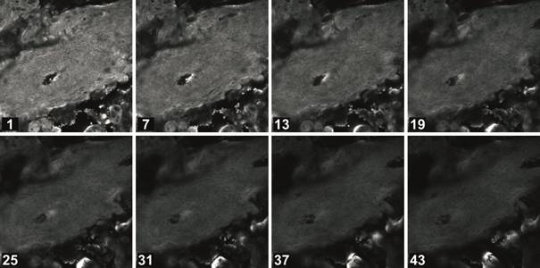
Fig. 1. Subset of a series of optical sections of a
human placenta captured by a confocal microscope. The distance between sections
in the subset is about 3 microns. The numbers in the figure
depict numerical order of optical sections in the full series.
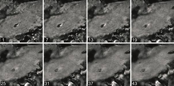
Fig. 2. Corrected subset
of a series of confocal optical sections of a human placenta.